
Diabetes Mellitus: Disease Management
Robert. S. Zimmerman
Published: October 2013
Definition
The microvascular complications of diabetes encompass long term complications of diabetes affecting small blood vessels. These classically have included retinopathy, nephropathy and neuropathy.Retinopathy is divided into 2 main categories. Non-proliferative retinopathy and proliferative retinopathy.
Non-proliferative retinopathy can be recognized by development of microaneurysms, venous loops, retinal hemorrhages, hard exudates and soft exudates.Proliferative retinopathy is defined as presence of new blood vessels with or without vitreous hemorrhage.Proliferative retinopathyrepresents a progression of non-proliferative retinopathy.
Diabetic nephropathyis defined as the presence of persistent proteinuria >0.5 g/24 hours.Overt nephropathyis characterized by progressive decline in renal function resulting in end stage renal disease.
Neuropathyis a heterogeneous condition that is associated with nerve pathology. The condition is classified according to the nerves affected. The classification of neuropathy includes focal, diffuse, sensory, motor and autonomic neuropathy.
Macrovascular complications of diabetes include coronary artery disease, stroke and peripheral vascular disease. Early macrovascular disease is associated with atherosclerotic plaque in vessels supplying blood to the heart, brain, limbs, and other organs. Late stages of macrovascular disease involve complete obstruction of these vessels which can include myocardial infarction (MI), stroke, claudication, and gangrene.
Prevalence
In type 1 diabetic patients, 13% have retinopathy at 5 years and 90% have retinopathy after 10 to 15 years. Approximately 25% of type 1 diabetics develop proliferative retinopathy after 15 years of diabetes.1 Type 2 diabetics taking insulin have a 40% prevalence of retinopathy at 5 years, while those on oral hypoglycemic agents have a 24% prevalence. By 15 to 19 years of diabetes, the rates increase to 84% and 53% respectively. Proliferative retinopathy develops in 2% of patients who have had type 2 diabetes for longer than 5 years, and in 25% of those who have had diabetes for 25 years or longer.2
The prevalence of nephropathy in diabetes has not been determined. Approximately 30% of patients with type I diabetes–and 5% to 10% of those with type 2 diabetes–become uremic.3 The prevalence of neuropathy, defined by loss of ankle jerk reflexes is 7% at 1 year, increasing to 50% at 25 years4 for both type 1 and type 2 diabetes. It has been estimated that 37% to 42% of all ischemic strokes in both African Americans and whites are attributable to the effects of diabetes alone or in combination with hypertension.5 The prevalence of heart disease or stroke in patients with diabetes is thought to be approximately 33.7% in men and 33.5% in women. The prevalence of peripheral vascular disease in patients with diabetes aged ≥ 30 years is 26%.6
Pathophysiology
Microaneurysm formation is the earliest manifestation of diabetic retinopathy. Microaneurysms may form due to the release of vasoproliferative factors, weakness in the capillary wall, or increased intra-luminal pressures. Microaneurysms can lead to vascular permeability. Vascular permeability in the macula can lead to macular edema and can threaten central vision. Obliteration of retinal capillaries can lead to intraretinal microvascular abnormalities (IRMAs). As capillary closure becomes extensive, intraretinal hemorrhages develop.
Proliferative retinopathy develops due to ischemia and release of vasoactive substances, such as vascular endothelial growth factor (VEGF), which stimulates new blood vessel formation as a progression of non-proliferative retinopathy. These vessels erupt through the surface of the retina and grow on the posterior surface of the vitreous humor. These vessels are very friable and can lead to vitreous hemorrhages. The vitreous humor can contract and lead to retinal detachment.
The pathophysiology of neuropathy is complex. Diabetes is associated with dyslipidemia, hyperglycemia, and low insulin and growth factor abnormalities. These abnormalities are associated with glycation of blood vessels and nerves. In addition, autoimmunity may affect nerve structure. Trauma and nerve entrapment can lead to structural nerve damage including segmental demyelination, axonal atrophy and loss, and progressive demyelination. These effects cause neuropathy. Several agents including laminin B2, immunoglobulin FI (IGFI) and II, nerve growth factor (NGF), insulin, and neurotrophin-3 (NT3) are potential growth factors that may restore nerve function.7
Diabetic nephropathy results from increased glomerular capillary flow that in turn results in increased extracellular matrix production and endothelial damage. This leads to increased glomerular permeability to macromolecules. Mesangial expansion and interstitial sclerosis ensues leading to glomerular sclerosis.
The macrovascular complications of diabetes result from hyperglycemia, excess free fatty acid, and insulin resistance. These cause increased oxidative stress, protein kinase activation and activation of the receptor for advanced glycation end products (RAGE). These factors act on the endothelium . First, decreased nitric oxide, increased endothelin, and increased angiotensin II cause vasoconstriction that results in hypertension and vascular smooth muscle cell growth. Second, decreased nitric oxide, activated nuclear factor-KB (NFKB), increased angiotensin II, and activation of activated protein-1 cause increased inflammation. This results in the release of chemokines, and cytokines, and expression of cellular adhesion molecules. Third, decreased nitric oxide, increased tissue factor, increased plasminogen activator inhibitor-1, and decreased prostacyclin result in thrombosis, hypercoagulation, platelet activation, and decreased fibrinolysis. All of these pathways lead ultimately to atherosclerosis, the cause of the macrovascular complications of diabetes.8
Signs and Symptoms
Symptoms of retinopathy are minimal until advanced disease ensues with loss or blurring of vision. Signs of non-proliferative retinopathy include microaneurysms, venous loops, retinal hemorrhages, hard exudates, and soft exudates. Proliferative retinopathy can include new vessels in the eyes or vitreous hemorrhage.
The earliest sign of nephropathy is hypertension. Development of hypertension often coincides with the development of microalbuminuria. As nephropathy worsens, patients can develop edema, arrhythmias associated with hyperglycemia, and/or symptoms related to renal failure.
Signs and symptoms of neuropathy are dependent on the type of neuropathy that develops. Most commonly, patients develop symptomatic distal polyneuropathy. Signs include depression or loss of ankle jerks and vibratory sensation, with hyperalgesia and calf pain in some patients. The deficit is in a stocking glove distribution. Wasting of the small muscle of the hands and feet can also occur.
Patients may present with focal neuropathies due either to mononeuritis or entrapment syndromes. These produce focal neurologic deficits confined to a single nerve. A rare but severe form of diabetic neuropathy is diabetic amyotrophy, which begins with pain followed by severe weakness and spreads from unilateral to bilateral. It resolves spontaneously in 18-24 months.
Patients with coronary artery disease can present with stable angina pectoris, unstable angina pectoris, or myocardial infarction. Many patients have unrecognizable symptoms and can present with dysrhythmias. Patients with cerebral vascular disease can present with a sudden onset of a focal neurologic deficit such as facial droop, hemiparesis, or isolated weakness of an arm or leg. Dizziness, slurred speech, gait difficulties, and visual loss can also be presenting symptoms. Stroke symptoms that last <24 hours are referred to as atransient ischemic event.
Peripheral vascular disease is recognized by exertional leg pain that can progress to rest pain and ischemic ulcers. Most cases are asymptomatic.
Screening
Patients with diabetes should be screened regularly for complications (Table 1).
Table 1: Microvascular complications in diabetes mellitus: screening and interventions
| Complication | Detection | Primary Prevention | Secondary Prevention |
|---|---|---|---|
| Retinopathy |
|
|
|
| Nephropathy | Urine micoalbumin |
|
|
| Neuropathy | Monofilament testing (see Figure 1)9 | Daily foot inspection |
|
ACEI, angiotensin-converting enzyme inhibitor; ARB, angiotensin receptor blocker; BP, blood pressure; PKC β, protein kinase C beta; VEGF, vascular endothelial cell growth factor.
*See elsewhere in the Disease Management Project: “Prevention and Treatment of Leg and Foot Ulcers in Diabetes Mellitus“)
Diabetic Retinopathy
Dilated eye examinations by ophthalmologists or optometrists should be performed within 5 years of onset in T1DM and at the time of diagnosis in T2DM, because the actual date of onset is unknown in T2DM.10,11 Follow-up examinations should be performed annually in patients with no or minimal background retinopathy. More frequent follow-up examinations should be performed in those who have more advanced retinopathy. Handheld ophthalmoscopy in the office may be able to detect diabetic retinopathy but offers limited view of the retina and difficulty detecting diabetic macular edema. Macular edema is a significant cause of vision loss in DM. It is easier to detect with binocular vision and, in difficult cases, IV fluorescein angiography and confocal microscopy are used. Technology is available for screening with fundus photographs obtained in the physician’s office and then read by an experienced reader. However, these methods are not yet sufficiently standardized to be used as routine screening tools.
Diabetic Nephropathy
The hallmark of early diabetic nephropathy is albumin excretion. Sensitive assays to detect very low levels of albumin, or microalbuminuria, have been available for many years.10,11 The simplest screening measure is a spot urine test adjusted for the urine creatinine level. Timed overnight collections and 24-hour collections may also be used. In general, microalbuminuria is defined as more than 30 mg albumin per gram of creatinine (spot urine test) or 30 to 299 mg per 24 hours and more than 300 mg/g creatinine (or 24 hours) as albuminuria. Serum creatinine determinations should be performed at least annually in patients with albuminuria; when estimated glomerular filtration rate (GFR) values are declining, more-specific measures of GFR (most commonly, creatinine clearance) should be used.
Peripheral Neuropathy
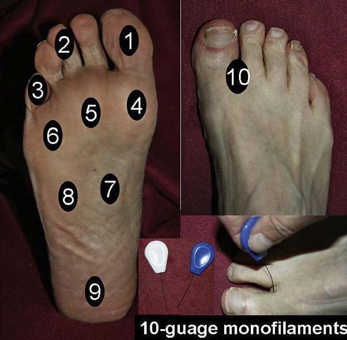
Monofilament testing in the office is the easiest way to test for the insensate foot.9,12-18 The 5.07-mm monofilament should be applied to the bottoms of the feet (Figure 1).9 Any loss of sensation is associated with an increased risk for ulcer formation. Any patient who has had a foot ulcer is at increased risk for additional foot ulcers. Patients should be instructed to examine their feet daily. Patients who have difficulty examining their feet should seek assistance, especially if the patient has impaired vision. The use of a mirror, such as a magnifying shaving mirror, can help the patient see the bottoms of his or her feet (see elsewhere in this section, “Prevention and Treatment of Leg and Foot Ulcers in Diabetes Mellitus“).
Coronary Heart Disease
Careful questioning about symptoms of ischemic coronary disease is still one of the most important ways to screen. Many patients with diabetes do not have typical exertional chest pain. Consequently, clinicians must ask about reduced exercise tolerance, dyspnea, or exercise-induced nausea.
Various studies have considered the issue of screening for coronary heard disease (CHD).10,1119, 20-23 The guidelines and individual recommendations are not entirely concordant. Whereas nearly every group suggests stress tests for patients with symptoms of CHD or electrocardiographic changes suggesting ischemia, recommendations on screening for asymptomatic disease are less consistent. The ADA considers that candidates for a screening cardiac stress test should include those with a history of peripheral or carotid occlusive disease; persons with a sedentary lifestyle, who are aged >55 years, and who plan to begin a vigorous exercise program; and those with 2 or more of the risk factors noted earlier.11 The American Association of Clinical Endocrinologists (AACE) guidelines state:
“Screening for asymptomatic coronary artery disease is an important consideration in patients with diabetes, but no data have shown that screening improves outcomes in this group and it is not recommended.”10
The AHA consensus group has provided a thoughtful approach to screening for CHD in patients with diabetes. They noted:
“Screening is defined here as the detection of disease in asymptomatic persons. Because screening tests are intended for widespread application, they should be rapid and inexpensive. In addition, to be useful, the results of testing should lead to a change in management, and the results of testing should improve outcome.”17
The American College of Cardiology (ACC)/AHA Guidelines for Exercise Testing give screening by exercise treadmill testing in patients with diabetes a data quality rating of IIb; that is, its usefulness or efficacy is less well established by evidence or opinion.23 They add that exercise testing “might be useful in people with heightened pretest risk.”
Most consensus statements and guidelines on diabetes and CHD have suggested that noninvasive cardiac testing be performed in patients with diabetes and 1 additional criterion: peripheral arterial disease, cerebrovascular disease, rest changes on the electrocardiogram (ECG), or the presence of ≥ 2 or more major coronary vascular disease (CVD) risk factors.
According to these guidelines, risk assessment begins with a medical history, including special attention to symptoms of atherosclerotic disease, such as angina, claudication, or erectile dysfunction. Electrocardiographic changes showing left ventricular hypertrophy and ST-T changes suggest increased cardiovascular risk. The ongoing DIAD study, which is designed to determine risk factors associated with clinically silent myocardial disease using stress tests with cardiac imaging, has suggested that the presence of neuropathy may be one of the most important predictors of cardiovascular risk.
It is not yet clear exactly how noninvasive testing changes risk management strategies in diabetes, because DM is already considered a coronary heart disease (CHD) risk equivalent. Thus, noninvasive testing should be targeted as much as possible to detect patients who might have CHD that is amenable to surgical intervention. Whereas noninvasive screening in asymptomatic patients might detect disease amenable to percutaneous intervention or coronary artery bypass grafting, the cost-effectiveness and determination of how much such screening affects long-term outcomes are still uncertain.
Careful attention to history of changes in exercise tolerance, atypical symptoms that suggest angina, or suggestive electrocardiographic abnormalities, are reasons for the clinician to consider stress testing. In addition, dyslipidemia, obesity, and hypertension, albuminuria, and a family history of CHD may be reasons to consider stress testing in patients who do not have clinical symptoms of CHD. This approach is most consistent with the AACE guidelines and should select patients at highest risk for CHD. In the absence of robust evidence, as noted by the AHA, physicians still need to make decisions about patients who might have silent myocardial disease.
Diagnosis
The diagnosis of nephropathy is initially based on development ofmicroalbuminuria. Microalbuminuria is defined as an albumin excretion rate 20 to 200 mcg/min. Because the average daily albumin excretion rate varies in normals and diabetics by 40%, it is recommended that 3 urine collections over several weeks be taken before this diagnosis is made. Overt nephropathy is defined as an albumin excretion rate >300 mg/24 hours. This is associated with a linear decline in GFR ranging from 0.1 to 2.4 mL/min/month. Stage 1 nephropathy is associated with a GFR >90 mg/24 hours. Stage 2 includes a mildly decreased GFR of 60 to 89 mg/24 hours. In stage 3 nephropathy, the GFR declines to 30 to 59 mg/24 hours, and in stage 4 GFR is 15 to 29 mg/24 hours. Stage 5 is end-stage nephropathy with a GFR <15 mg/24 hours.
Diagnosis of retinopathy is based on the findings of eye exams as discussed under “Definition” above.
The diagnosis of neuropathy is based on finding focal (individual root) or diffuse (entire limb) involvement. Findings can be asymmetric ( mononeuritis multiplex) or symmetric conforming to a distal-to-proximal gradient of involvement (most common). Electrodiagnostic studies can confirm peripheral nerve disease and define the pattern of disease. Autonomic neuropathy is diagnosed in patients with gastroparesis or orthostatic hypotension.
The diagnosis of coronary artery disease can be confirmed by several diagnostic and imaging studies. A resting 12-lead ECG is not sensitive enough to identify disease in patients with stable angina. Cardiovascular stress testing can be assessed with ECG assessment during exercise, dobutamine, dipyridamole, or adenosine. Echocardiography can further enhance the sensitivity of the test. Alternatively nuclear stress testing with thallium 201 or technetium 99m in association with dipyridamole or adenosine can be used. Significant CAD is identified by relative hypoperfusion in peak stress images. Confirmation of the coronary artery disease is made by coronary arteriography.
The diagnosis of a stroke is based on a patient developing symptoms as above and is confirmed by a CT scan or MRI. CT angiography can be used to identify the location of vascular occlusion and assess for salvageable brain tissue.
The diagnosis of peripheral arterial disease is diagnosed by determining the ankle brachial index (ABI). This is the ratio of the Doppler determined systolic ankle pressure over the systolic brachial pressure. ABI <0.9 has a sensitivity of 95% and a specificity of 100% in detecting PAD. An ABI >1.4 reflects calcified arteries. It is associated with increased risk of foot ulcers and CVD. If revascularization is being considered other tests including duplex ultrasonography, MR angiography, and CT angiography can be used to determine specific sites of surgical intervention.
Therapy / Management
Lifestyle Treatment
Medical Nutrition Treatment
Guidelines for medical nutrition therapy11,24-28 have been established by the ADA and are summarized in (Figure 2). The primary focus of these guidelines is targeted to improve outcomes including glycemic control, weight reduction (as appropriate), blood pressure control, and a favorable lipid profile. There is clear evidence that excess saturated fat in the diet has a detrimental effect on lipid profiles, and therefore restriction of saturated fat is recommended. The data supporting absolute restriction of carbohydrates are not robust, so the ADA guidelines allow flexibility in intake of carbohydrates and nonsaturated fat. Separate guidelines have been published about the carbohydrate content and composition of the diet.29
Figure 2. Goals of medical nutrition therapy for all persons with diabetes24
Attain and maintain optimal metabolic outcomes, including the following:
|
| Improve health through healthy food choices and physical activity. |
| Address individual nutritional needs, taking into consideration personal and cultural preferences and lifestyle while respecting the individual’s wishes and willingness. Specific Situations Children and Adolescents with Type 1 Diabetes |
| Provide adequate energy to ensure normal growth and development. |
| Integrate insulin regimens into usual eating and physical activity habits. Children and Adolescents with Type 2 Diabetes |
| Facilitate changes in eating and physical activity habits that reduce insulin resistance and improve metabolic status. Pregnant and Lactating Women |
| Provide adequate energy and nutrients needed for optimal outcomes. Older Adults |
| Provide for the nutritional and psychosocial needs of aging adults. Persons Treated with Insulin or Insulin Secretagogues |
| Provide self-management education for treatment (and prevention) of hypoglycemia, acute illnesses, and exercise-related blood glucose problems. Persons at Risk for Diabetes |
| Decrease risk by encouraging physical activity and promoting food choices that facilitate moderate weight loss or at least prevent weight gain. |
The most important variable in prandial glycemic excursion is total carbohydrate intake. Low glycemic index foods consumed alone result in lower prandial glucose excursion than do high glycemic index foods. However, in the context of a mixed meal, differences between low and high glycemic index foods are attenuated. The amount29-31 and source31,32 of carbohydrates are important determinants of postprandial glucose levels. The relative effects of each have been recently studied. Brand-Miller and colleagues33,34 have reported that they analyzed the relative impact of the glycemic index and total carbohydrate content of individual foods on glycemic load–the product of glycemic index and total grams of carbohydrate–using linear regression analysis. Carbohydrate content (total grams) alone explained 68% of the variation in glycemic load, and the glycemic index of the food explained 49%. When total carbohydrate and glycemic index were both included in the regression analysis, the glycemic index accounted for 32% of the variation.
Restriction of alcohol and sodium is generally advised. Nutritional supplements are not necessary in patients who are otherwise consuming a well-balanced diet. Many recommendations for weight management propose restriction of calories based on the degree of obesity and propose 30 to 45 minutes of exercise 3 to 5 days a week. Exercise is an important component of any regimen for weight reduction and glycemic control. Other nutritional guidelines for patients with diabetes are generally consistent with the ADA guidelines.10,19,26,35-47
Exercise
Guidelines for exercise have not always been specific with regard to exact exercise prescriptions, especially regarding aerobic and resistance exercises.10,11,14,29,48 The commonly proposed recommendation that 150 minutes of moderate-intensity (or 90 minutes of vigorous) aerobic exercise per week is generally the amount of exercise required to achieve benefits on glycemic control and reduce CHD risk and has been supported by ADA/American Heart Association (AHA) recommendations.27,28
Regular exercise is encouraged, but complications of diabetes need to be taken into account. Injury to patients with loss of sensation in their feet is a limitation for weight-bearing exercise. Because of risk of CHD in patients with diabetes, appropriate screening for CHD should be performed before patients engage in an exercise program.10,11,19,48-50 Benefits of exercise include weight control and improved glycemic control, often due to reduced insulin resistance.
The optimal approach to the microvascular complications of diabetes is prevention. The 2 main approaches to preventing retinopathy and nephropathy are intensive glycemic control and aggressive control of hypertension. Intensive glycemic control has been the most effective approach to preventing neuropathic complications of diabetes.
Glycemic Control
The Wisconsin Epidemiologic Study1,2,51-55 demonstrated that in both diabetics aged <30 years and in those aged >30 years treated with oral hypoglycemic agents or insulin, baseline HbA1C level correlated with the incidence of retinopathy, progression of retinopathy, and progression of proliferative retinopathy.
The Diabetes Control and Complications Trial56 (DCCT) enrolled 1,441 people with type 1 diabetes. Of these, 726 had no retinopathy, normal albumin excretion, and diabetes for <5 years; 715 had mild-to-moderate background retinopathy with normal or micro-albuminuria at baseline.
The subjects received intensive therapy or conventional treatment. The intensive treatment was either given with insulin pumps or multiple daily injections (3 or more injections per day.) The insulin dosage was guided by self-monitoring of blood sugar 3 or 4 times per day. The participants were seen every month.
The conventional group received no more than 2 shots per day. Urine and blood sugars were monitored no more than 2 times per day. They had clinic visits every 2 or 3 months over an average of 6.5 years. The average hemoglobin A1C (HbA1C) was 9.1% in the conventional group and 7.2% in the intensively treated group throughout the study. Risk reduction was 70% for clinically important sustained retinopathy, 56% for laser photo coagulation, 60% for sustained micro-albuminuria, 54% for clinical grade nephropathy, and 64% for clinical neuropathy. Four years after the close of the DCCT, HbA1C levels in the 2 groups narrowed to 8.2% in the conventional treatment group and 7.9% in the intensive treatment group. Retinopathic events including proliferative retinopathy, macular edema, and need for laser therapy were 74%, 77%, and 77% lower in the intensively treated group. The incidence of micro-albuminuria was 53% lower and the incidence of albuminuria 86% lower in the intensively treated group.57
The Kumamato Trial58 included 102 patients with type 2 diabetes and showed that intensive therapy with multiple daily injections (preprandial, regular, and bedtime intermediate acting insulin) compared with once or twice daily insulin injections resulted in a decrease in HbA1C from 9.4% to 7.1%. Two-step progression of retinopathy decreased by 69%, nephropathy progression decreased by 70%, and nerve conduction velocities improved.
The United Kingdom Prospective Diabetes Study59,60 evaluated 5,102 patients with type 2 diabetes. Patients were treated with sulphonylureas or insulin in 1 study and another group was compared to these groups using metformin in a second study. The study subjects maintained an average HbA1C of 7.9% in the conventional treatment group compared to 7% in the intensive treatment group. There was a 27% risk reduction for retinal photo coagulation at 12 years, 33% risk reduction at 12 years for micro-albuminuria, and 74% risk reduction for doubling of creatinine at 12 years.
Blood pressure control has been shown to reduce the risk for both retinopathy and nephropathy. The Hypertension and Diabetes Study61,62 was part of the UKPDS Study. There were 1,148 patients with type 2 diabetes and coexisting hypertension that were studied. Tight control subjects were given a blood pressure goal of <150/85 mm Hg on treatment. Most patients were treated with captopril or atenolol. The control group was given a blood pressure goal of <180/105 mm Hg.
On average, patients in the tight control group maintained an average blood pressure of 144/82 mm Hg, while those in the control group averaged 154/87 mm Hg. Tight control resulted in a 35% reduction in retinal photo coagulation (P <0.025), a 34% reduction in 2-step deterioration of retinopathy, and a 47% risk reduction in 3-deterioration in the ETDRS chart (P <0.005) over 7.5 years.
The Euclid Trial,63 which included 354 normotensive patients with type 1 diabetes aged 20 to 59 years, demonstrated that lisinopril treatment resulted in a 50% reduction in retinopathy progression, 73% reduction in 2-grade retinopathy progression, and an 82% reduction in development of proliferative retinopathy.
Several studies have assessed the effects of blood pressure control on nephropathy in patients with type 1 and type 2 diabetes have been performed assessing effects. Parving64 demonstrated that blood pressure control in patients with diabetes with nephropathy decreased albumin excretion rate by 50% and the rate of decline of GFR from 0.29 to 0.1 mL/min/month.
A recent meta analysis65 involving multiple studies66-76 demonstrated that ACE inhibitors can delay progression to overt nephropathy by 62% in patients with type 1 diabetes with micro-albuminuria. Many of these patients also experienced decreased albumin excretion rates. No studies in type 1 patients have shown that starting ACE inhibitors when the albumin excretion rate is normal delays the development of micro-albuminuria.
Lewis77 studied 409 patients with type 1 diabetes with overt nephropathy (protein excretion >500 mg/d and creatinine <2.5 mg/dL). Creatinine doubled in 12.1% of the patients receiving captopril and in 21.3% of patients receiving placebo (a 48% reduction in risk).
Several studies in patients with type 2 diabetes with micro-albuminuria─with or without hypertension─have found that ACE inhibitors delay progression to overt nephropathy, decrease the albumin excretion rate, and diminish the decline in GFR.78-85 One study demonstrated that in type 2 diabetics who were normotensive and normoalbuminuric that enalapril attenuated the increase in the albumin excretion rate and decreased the likelihood of development of micro-albuminuria (a 12.5% risk reduction).86
Several studies87-90 have shown that in patients with type 2 diabetes, there is a slowing of progression of micro-albuminuria to overt nephropathy when angiotensin II receptor blockers are administered. Based on these studies, the American Diabetes Association (ADA) has recommended11 a preprandial plasma glucose goal of 70 to 130 mg/dL and a postprandial glucose goal of <180 mg/dL. Normal HbA1C is <6% and the ADA goal is <7%.
The ADA target for blood pressure is <130/80 mm Hg. The American Association of Clinical Endocrinology (AACE) recommends preprandial glucose targets of <110, post-prandial, <140, and an HbA1C <6.5%.10 The AACE also recommends a blood pressure goal of <130/85 mm Hg. HbA1C measurements are suggested every 3 months. Blood sugar testing in type 1 diabetics or pregnant women with diabetes is recommended at least 3 times a day.
The frequency of glucose monitoring for type 2 diabetics is not known but should be sufficient to facilitate achievement of the glucose goals. In hypertensive patients with micro-albuminuria or albuminuria, ACE inhibitors or angiotensin II receptor blockers should be strongly considered. The UKPDS found that intensive blood pressure control decreased microvascular complications by 37% with both ACE inhibitors and beta blockers.62
Patients with type 1 diabetes should have an initial dilated and comprehensive eye exam within 3 to 5 years of the onset of diabetes. Patients with type 2 diabetes should have an eye exam shortly after diagnosis. Both Type 1 and Type 2 diabetics should have subsequent eye exams annually, which should be performed by an ophthalmologist or optometrist knowledgeable and experienced in diagnosing retinopathy.
Once retinopathy is established, the best treatment to prevent blindness in those with proliferative retinopathy is laser photo-coagulation.91-95 The Diabetic Retinopathy Study found that a 50% reduction in severe visual loss could be achieved by treating eyes with neovascularization associated with vitreous hemorrhage, or neovascularization on or near the optic disc, and eyes with proliferative retinopathy or very severe non-proliferative retinopathy.91-95 If vitreous hemorrhage occurs and does not resolve, vitrectomy can be performed to restore vision.
Early nephropathy is associated with micro-albuminuria, hypertension, and possible elevation in creatinine. First-line therapy is directed toward controlling hypertension. Generally, ACE inhibitors are agents of first choice. If patients develop a cough, angioreceptor blockers have shown similar efficacy at decreasing micro-albuminuria, lowering blood pressure, and preventing worsening renal function. Some calcium channel blockers (diltiazem and verapamil) have been shown to decrease microalbuminuria and may be added to the above medications if necessary. If creatinine increases above 2 or 3 mg/dL, ACE inhibitors should be avoided because overt renal failure can result. If renal failure develops, treatment with dialysis or kidney transplant should be considered.
The Diabetes Control and Complications trial found some improvement in neuropathy with intensive diabetes control. If this is not successful, further treatment of neuropathy should focus on analgesia. The most common neuropathy is bilateral distal polyneuropathy. Increasing doses of tricyclic antidepressants, gabapentin, phenytoin, carbamazepine, and benzodiazepines have been used with varying degrees of success. Gastroparesis is treated with metoclopramide.
Management of Coronary Heart Disease Risk
Dyslipidemia
Guidelines for the management of dyslipidemia have been published by the National Cholesterol Education Program (NCEP) and by several expert panels since 1988. They include the AACE, ACP, ADA, ACC, and AHA (Table 2). The guidelines are generally consistent in recommending aggressive lipid-lowering management in diabetes, which is considered a coronary risk equivalent.
Table 2: Goals for Risk Factor Management in Patients With Diabetes19
| Risk Factor | Goal of Therapy | Recommending Body |
|---|---|---|
| Cigarette smoking | Complete cessation | ADA |
| Blood pressure | <130/85 mm Hg | JNC VI (NHLBI) |
| <130/80 mm Hg | ADA | |
| LDL cholesterol level | <100 mg/dL | ATP III (NHLBI), ADA |
| Triglyceride level 200-499 mg/dL |
Non-HDL cholesterol level <130 mg/dL |
ATP III (NHLBI) |
| HDL cholesterol level <40 mg/dL |
Raise HDL (no set goal) |
ATP III (NHLBI) |
| Prothrombotic state | Low-dose aspirin therapy (patients with CHD and other risk factors) |
ADA |
| Glucose | HbA1c <7% | ADA |
| Overweight and obesity (BMI ≥ 25 kg/m2) |
Decrease BMI | OEI (NHLBI) |
| Physical inactivity | Exercise prescription depending on patient’s status |
ADA |
| Adverse nutrition | See text | ADA, AHA, and NHLBI’s ATP III, OEI, and JNC VI |
ADA, American Diabetes Association; AHA, American Heart Association; ATP III, National Cholesterol Education Program Adult Treatment Panel III; BMI, body mass index; CHD, coronary heart disease; HDL, high-density lipoprotein; JNC VI, Sixth Report of the Joint National Committee on Prevention, Evaluation, and Treatment of High Blood Pressure; LDL, low-density lipoprotein; NHLBI, National Heart, Lung, and Blood Institute; OEI, Obesity Education Initiative Expert Panel on Identification, Evaluation, and Treatment of Overweight and Obesity in Adults.
Physicians should note that not all patients with diabetes have a 20% risk of a cardiac event over a 10-year period as determined by the UKPDS risk engine,39 so some discretion may be used with the guidelines. The proposed LDL cholesterol level targets are as follows:
- The LDL cholesterol level <100 mg/dL for any patient with DM.
- If the LDL cholesterol level is <100 mg/dL, but triglyceride (and very LDL [VLDL] cholesterol) levels are elevated, then the non–high-density lipoprotein (HDL) cholesterol level should be <130 mg/dL.
- The optional guidelines for patients at very high risk, such as diabetic patients with a prior myocardial infarction (MI), are an LDL cholesterol level <70 mg/dL (and non-HDL cholesterol level <100 mg/dL).
- Patients who have an LDL cholesterol level <100 mg/dL without medication should be treated to achieve a >30% reduction in the LDL cholesterol level.
These guidelines were developed based on findings from lipid-lowering trials that included diabetic patients and were confirmed by subsequent trials.
Post-hoc analyses of diabetic patients who were included in lipid-lowering trials have supported the notion that these patients have comparable relative reductions (or perhaps greater absolute reductions) in the risk for CHD events than their nondiabetic counterparts. These data have been summarized as part of the ACP guidelines.39The ADA and AHA guidelines6,7,11,27-29,55,56,96,97 have suggested an LDL cholesterol level target of <100 mg/dL for patients with diabetes and an optional target of <70 mg/dL for patients with DM who already have CHD. This recommendation is based on several clinical trials including, the HPS, ASCOT-LLA, and CARDS trials.98-101The CARDS trial, in 2838 patients with T2DM, showed a 37% reduction in cardiovascular events, with a mean in-trial LDL cholesterol level of approximately 80 mg/dL in the atorvastatin group compared with 119 mg/dL in the placebo group. The HPS and CARDS studies have shown favorable effects in diabetic patients whose LDL cholesterol levels were <100 mg/dL. In addition, these guidelines have recommended that in patients with elevated triglyceride levels, and a corresponding increase in VLDL cholesterol levels, that the non-HDL cholesterol value (LDL plus VLDL cholesterol level) be set at 30 mg/dL higher than the LDL target–that is, a non-HDL cholesterol level <130 mg/dL, with an optional target of <100 mg/dL.
The FIELD trial was designed to assess the effects of fenofibrate vs. placebo on cardiovascular disease events in 9795 T2DM subjects.102-103 The difference in total cardiovascular events was 11% (P = .035) and in MI plus CHD death it was 11% (P = .16). This trial was confounded by very high levels of statin drop-in, especially in the placebo arm. The ACCORD trial did not find that fenofibrate decreased cardiac events in the entire population but did so in those with high triglycerides and low HDL cholesterol.
Hypertension and Renin-Angiotensin-Aldosterone System Blockade
Blood pressure control has a greater effect on reducing the risk for stroke than it does for reducing the risk for MI. The largest blood pressure trials in diabetic patients have demonstrated favorable effects on reduction in CVD. Current clinical guidelines recommend BP targets of 130/80 (or 130/85) mm Hg.10,11,102,104,107 Few clinical trials have actually achieved these goals, but there does not appear to be any risk in reaching these targets. Multidrug regimens (often 3 or more drugs) are usually required. Based on several studies (especially HOPE and the EUROPA study, which demonstrated favorable cardiovascular effects with the ACE inhibitors ramipril and perindopril, respectively, in diabetic cohorts), these agents should be considered part of initial therapy in hypertensive T2DM subjects.105,108 ONTARGET (N = 25,620; 38% with diabetes) compared ramipril to telmisartan and to the combination of both drugs on CVD outcomes. There were no differences among the 3 arms.109 The beneficial effects could not be entirely attributed to blood pressure reduction in these trials. The ACCORD trial showed no benefit in decreasing BP <120 mm Hg versus <140 mm Hg.
Aspirin
Aspirin (ASA) therapy is recommended by the ADA guidelines, and other, for primary prevention in patients with diabetes.11,110 In men aged ≥50 years and in women aged ≥60 years with type 1 or type 2 diabetes plus an additional risk factor, ASA should be used for secondary prevention. ASA should be used in combination with clopidogrel for up to 1 year in these patients following acute coronary syndrome.
Smoking
All the DM- and CHD-related guidelines recommend smoking cessation.
Glycemic Control
Intervention trials have shown a somewhat modest a relationship between glycemic control and CHD risk. In the UKPDS trial, a delta HbA1c value of 0.9% was associated with a 14% reduction in the risk for MI (P = .052) in the intention to treat analyses and a 16% reduction for every 1% HbA1c level change as a post hoc observational analysis.60,111,112 The metformin arm in obese patients in the UKPDS demonstrated a 39% reduction in MI compared with the conventional arm (P = .010).59 ACCORD (N = 10,251) and ADVANCE (N = 11,140) did not demonstrate beneficial effects of intensive control (HbA1c <7.0%) on CVD events.113,114
In the DCCT/EDIC study, there was no statistically significant reduction in CHD risk at the end of the DCCT. This was expected, because the trial included a population at low risk for CHD at randomization. However, a 42% reduction (P = .016) in risk of any cardiac event during the duration of the DCCT/EDIC study indicates that the annualized effect of glycemic control on CHD risk is less than that generally associated with other interventions, especially lipid lowering.115
Two recent studies have suggested that patients with a BMI ≥ 35 may benefit from gastric bypass surgery as treatment for obesity and diabetes. Mingrone et al116 found that in patients aged 30 to 60 years, with BMI ≥35, 95% of patients treated with biliopancreatic diversion, and 75% of those treated with gastric bypass, had remission of diabetes. Schauer et al117 found that 42% of obese patients with gastric bypass, 37% obese patients with sleeve gastrectomy, and 12% of medically managed patients achieved HbA1c ≤6% at 1 year.
Outcomes
A patient with diabetes should be referred to an endocrinologist if targets for glycemic control cannot be achieved or if the patient is experiencing significant hypoglycemia. It is important to refer early in order to help patients avoid long-term complications. Patients who develop complications should be referred to an endocrinologist to see if glycemic control can be improved, or simply to treat the complications.
Summary
The watchword of the ADA several years ago was “diabetes is serious.” Careful screening for complications, including retinopathy, nephropathy, and neuropathy clearly are associated with opportunities to reduce the risk for disease progression. Aggressive interventions with glycemic control, as well as management of lipids and blood pressure, seem to have favorable effects on many complications of diabetes. Aspirin therapy also reduces the risk for CHD risk in patients with DM. These screening and intervention strategies are supported by robust observational and intervention trial data and, in turn, are endorsed by the various organizations that have written disease management guidelines.
Summary
- Diabetes mellitus is a leading cause of blindness, end-stage renal disease, and nontraumatic lower extremity amputations.
- Diabetes mellitus increases the risk for coronary heart disease by 2- to 5-fold.
- Glycemic control is associated with a reduced risk for the microvascular and neuropathic complications of diabetes mellitus.
- Treatment of CHD risk factors, especially dyslipidemia, is associated with a reduced risk for CHD.
- Early detection of microvascular and neuropathic complications and implementation of appropriate treatment strategies, such as laser therapy (retinopathy), use of ACE inhibitors and ARBs (nephropathy), and proper footwear (neuropathy), will reduce the risk for adverse outcomes from these complications.
Suggested Readings
- Adler AI, Stratton IM, Neil HA, et al: Association of systolic blood pressure with macrovascular and microvascular complications of type 2 diabetes (UKPDS 36): Prospective observational study. BMJ. 2000;321:412-419.
- American Diabetes Association. Standards of medical care in diabetes-2007. Diabetes Care. 2007;30:(Suppl 1)S4-S41.
- Bantle JP, Wylie-Rosett J, Albright AL, et al: Nutrition recommendations and interventions for diabetes-2006: A position statement of the American Diabetes Association. Diabetes Care. 2006;29:2140-2157.
- Buse JB, Ginsberg HN, Bakris GL, et al: Primary prevention of cardiovascular diseases in people with diabetes mellitus: A scientific statement from the American Heart Association and the American Diabetes Association. Diabetes Care. 2007;30:162-172.
- Diabetes Control and Complications Trial Research Group. The relationship of glycemic exposure (HbA1c) to the risk of development and progression of retinopathy in the Diabetes Control and Complications Trial. Diabetes. 1995;44:968-983.
- Diabetes Control and Complications Trial/Epidemiology of Diabetes Interventions and Complications Research Group. Retinopathy and nephropathy in patients with type 1 diabetes four years after a trial of intensive therapy. N Engl J Med. 2000;342:381-389.
- Gibbons RJ, Balady GJ, Beasley JW, et al: ACC/AHA guidelines for exercise testing: Executive summary. A report of the American College of Cardiology/American Heart Association Task Force on Practice Guidelines (Committee on Exercise Testing). Circulation. 1997;96:345-354.
- Grundy SM, Cleeman JI, Merz CN, et al: Implications of recent clinical trials for the National Cholesterol Education Program Adult Treatment Panel III guidelines. Arterioscler Thromb Vasc Biol. 2004;24:e149-e161.
- Stratton IM, Adler AI, Neil HA, et al: Association of glycaemia with macrovascular and microvascular complications of type 2 diabetes (UKPDS 35): Prospective observational study. BMJ. 2000;321:405-412.
- Strippoli GF, Craig M, Deeks JJ, et al: Effects of angiotensin-converting enzyme inhibitors and angiotensin II receptor antagonists on mortality and renal outcomes in diabetic nephropathy: Systematic review. BMJ. 2004;329:828.
References
- Klein R, Klein BEK, Moss SE, Davis MD, DeMets DL. The Wisconsin Epidemiologic Study of Diabetic retinopathy: II. Prevalence and risk of diabetic retinopathy when age at diagnosis is less than 30 years. Arch Ophthalmol. 1984;102:520-526.
- Klein R, Klein BEK, Moss SE, Davis MD, DeMets DL. The Wisconsin Epidemiologic Study of Diabetic retinopathy: II. Prevalence and risk of diabetic retinopathy when age at diagnosis is less than 30 years. Arch Ophthalmol. 1984;102:527-532.
- Friedman EA. Diabetic Renal Disease. In Rifkin H, Porte D, Eds. Diabetes Mellitus/Theory and Practice. 1990;684-709.
- Vinik AI, Mitchell BD, Leichter SB, Wagner AL, O’Brian IT, Georges LP. Epidemiology of the complications of diabetes. In: Leslie RDG, Robbins DC, eds. Diabetes: Clinical Science in Practice. Cambridge: Cambridge University Press, 1995:221.
- Kissela BM, Khoury J, Kleindorfer D, et al. Epidemiology of ischemic stroke in patients with diabetes: the greater Cincinnati/Northern Kentucky Stroke Study. Diabetes Care. 2005;28(2):355-359.
- Melton LJ 3rd, Macken KM, Palumbo PJ, Elveback LR. Incidence and prevalence of clinical peripheral vascular disease in a population-based cohort of diabetic patients. Diabetes Care. 1980;3:650-654.
- Vinik AI, Pittenger GL, McNitt P, Stansberry KB Diabetic Neuropathies: An overview of clinical aspects, pathogenesis, and treatment. LeRoith D, Taylor SI, Olefsky JM. Eds. A Fundamental and Clinical Text, 2nd Ed, 1990;911-934.
- Bechman, JA, Creager MA, Libby P. Diabetes and atherosclerosis: epidemiology, pathophysiology, and management. JAMA. 2002;287:2570-2581.
- Armstrong DG, Lavery LA. Diabetic foot ulcers: Prevention, diagnosis and classification. Am Fam Physician. 1998;57:1325-1328.
- American Association of Clinical Endocrinologists. The American Association of Clinical Endocrinologists medical guidelines for clinical practice for developing a comprehensive care plan. Endocr Pract. 2011;17(Suppl 2):1-53.
- American Diabetes Association. Standards of medical care in diabetes–2012. Diabetes Care. 2012,35;(Suppl 1):S11-S63.
- Armstrong DG, Lavery LA, Vela SA, et al: Choosing a practical screening instrument to identify patients at risk for diabetic foot ulceration. Arch Intern Med. 1998;158:289-292.
- Armstrong DG. The 10-g monofilament: The diagnostic divining rod for the diabetic foot? Diabetes Care. 2000;23:887.
- Boulton AJ, Vinik AI, Arezzo JC, et al: Diabetic neuropathies: A statement by the American Diabetes Association. Diabetes Care. 2005;28:956-962.
- Mayfield JA, Reiber GE, Sanders LJ, et al: Preventive foot care in diabetes. Diabetes Care. 2004; 27:(Suppl 1)S63-S64.
- Perkins BA, Olaleye D, Zinman B, Bril V. Simple screening tests for peripheral neuropathy in the diabetes clinic. Diabetes Care. 2001;24:250-256.
- Vinik AI. Diagnosis and management of diabetic neuropathy. Clin Geriatr Med. 1999;15:293-320.
- Wunderlich RP, Armstrong DG, Husain K, Lavery LA. Defining loss of protective sensation in the diabetic foot. Adv Wound Care. 1998;11:123-128.
- Grundy SM, Howard B, Smith S Jr, et al: Prevention Conference VI: Diabetes and Cardiovascular Disease: Executive summary: Conference proceeding for health care professionals from a special writing group of the American Heart Association. Circulation. 2002;105:2231-2239.
- Gibbons RJ, Balady GJ, Beasley JW, et al: ACC/AHA guidelines for exercise testing: Executive summary. A report of the American College of Cardiology/American Heart Association Task Force on Practice Guidelines (Committee on Exercise Testing). Circulation. 1997;96:345-354.
- Heller GV. Evaluation of the patient with diabetes mellitus and suspected coronary artery disease. Am J Med. 2005;118:(Suppl 2)9S-14S.
- Lebovitz HE, Austin MM, Blonde L, et al: ACE/AACE consensus conference on the implementation of outpatient management of diabetes mellitus: Consensus conference recommendations. Endocr Pract. 2006;12:(Suppl 1)6-12.
- Smith SC Jr, Allen J, Blair SN, et al: AHA/ACC guidelines for secondary prevention for patients with coronary and other atherosclerotic vascular disease: 2006 update: Endorsed by the National Heart, Lung, and Blood Institute. Circulation. 2006;113:2363-2372.
- Bantle JP, Wylie-Rosett J, Albright AL, et al: Nutrition recommendations and interventions for diabetes–2006: A position statement of the American Diabetes Association. Diabetes Care. 2006;29:2140-2157.
- Franz MJ, Horton ES Sr, Bantle JP, et al: Nutrition principles for the management of diabetes and related complications. Diabetes Care. 1994;17:490-518.
- Franz MJ, Bantle JP, Beebe CA, et al: Evidence-based nutrition principles and recommendations for the treatment and prevention of diabetes and related complications. Diabetes Care. 2003;26:(Suppl 1)S51-S61.
- Buse JB, Ginsberg HN, Bakris GL, et al: Primary prevention of cardiovascular diseases in people with diabetes mellitus: A scientific statement from the American Heart Association and the American Diabetes Association. Diabetes Care. 2007;30:162-172.
- Buse JB, Ginsberg HN, Bakris GL, et al: Primary prevention of cardiovascular diseases in people with diabetes mellitus: A scientific statement from the American Heart Association and the American Diabetes Association. Circulation. 2007;115:114-126.
- Sheard NF, Clark NG, Brand-Miller JC, et al: Dietary carbohydrate (amount and type) in the prevention and management of diabetes: A statement by the American Diabetes Association. Diabetes Care. 2004;27:2266-2271.
- Gannon MC, Nuttall FQ, Westphal SA, et al: Effects of dose of ingested glucose on plasma metabolite and hormone responses in type II diabetic subjects. Diabetes Care. 1989;12:544-552.
- Jenkins DJ, Wolever TM, Taylor RH, et al: Glycemic index of foods: A physiological basis for carbohydrate exchange. Am J Clin Nutr. 1981;34:362-366.
- Brand JC, Nicholson PL, Thorburn AW, Truswell AS. Food processing and the glycemic index. Am J Clin Nutr. 1985;42:1192-1196.
- Brand-Miller JC, Wolever TM. The use of glycaemic index tables to predict glycaemic index of breakfast meals. Br J Nutr. 2005;94:133-134.
- Mendosa R. Glycemic load values. Am J Clin Nutr. 2003;77:994-995.
- Clark MJ Jr, Sterrett JJ, Carson DS. Diabetes guidelines: A summary and comparison of the recommendations of the American Diabetes Association, Veterans Health Administration, and American Association of Clinical Endocrinologists. Clin Ther. 2000;22:899-910.
- Grundy SM. Approach to lipoprotein management in 2001 National Cholesterol Guidelines. Am J Cardiol. 2002;90:11i-21i.
- Krauss RM, Eckel RH, Howard B, et al: AHA Dietary Guidelines: Revision 2000: A statement for health care professionals from the Nutrition Committee of the American Heart Association. Circulation. 2000;102:2284-2299.
- Lichtenstein AH, Appel LJ, Brands M, et al: Diet and lifestyle recommendations revision 2006: A scientific statement from the American Heart Association Nutrition Committee. Circulation. 2006;114:82-96.
- Snow V, Aronson MD, Hornbake ER, et al: Lipid control in the management of type 2 diabetes mellitus: A clinical practice guideline from the American College of Physicians. Ann Intern Med. 2004;140:644-649.
- American Association of Diabetes Educators. AADE position statement: Medical nutrition therapy for people with diabetes mellitus. Diabetes Educ. 1995;21:17-18.
- American Association of Diabetes Educators. Position statement. Diabetes education and public health. Diabetes Educ. 2000;26:607-609.
- Kannel WB, McGee DL. Diabetes and cardiovascular disease. The Framingham study. JAMA. 1979;241:2035-2038.
- Krauss RM, Eckel RH, Howard B, et al: AHA Dietary Guidelines: Revision 2000: A statement for health care professionals from the Nutrition Committee of the American Heart Association. Circulation. 2000;102:2284-2299.
- Moss SE, Klein R, Klein BE, Meuer SM. The association of glycemia and cause-specific mortality in a diabetic population. Arch Intern Med. 1994;154:2473-2479.
- Diabetes Control and Complications Trial/Epidemiology of Diabetes Interventions and Complications Research Group. Epidemiology of Diabetes Interventions and Complications (EDIC). Design, implementation, and preliminary results of a long-term follow-up of the Diabetes Control and Complications Trial cohort. Diabetes Care. 1999;22:99-111.
- Diabetes Control and Complications Trial/Epidemiology of Diabetes Interventions and Complications Research Group. Retinopathy and nephropathy in patients with type 1 diabetes four years after a trial of intensive therapy. N Engl J Med. 2000;342:381-389.
- The Diabetes Control and Complications Trial/Epidemiology of Diabetes Interventions and Complications Research Group. Sustained effect of intensive treatment of type 1 diabetes mellitus on development and progression of diabetic nephropathy: The Epidemiology of Diabetes Interventions and Complications (EDIC) study. JAMA. 2003;290:2159-2167.
- Zinman B, Ruderman N, Campaigne BN, et al: Physical activity/exercise and diabetes. Diabetes Care. 2004;27:(Suppl 1)S58-S62.
- Gibbons RJ, Balady GJ, Beasley JW, et al: ACC/AHA Guidelines for Exercise Testing. A report of the American College of Cardiology/American Heart Association Task Force on Practice Guidelines (Committee on Exercise Testing). J Am Coll Cardiol. 1997;30:260-311.
- Lebovitz HE, Austin MM, Blonde L, et al: ACE/AACE consensus conference on the implementation of outpatient management of diabetes mellitus: Consensus conference recommendations. Endocr Pract. 2006;12:(Suppl 1)6-12.
- Klein R. Hyperglycemia and microvascular and macrovascular disease in diabetes. Diabetes Care. 1995;18:258-268.
- Klein R, Klein BE, Moss SE. Epidemiology of proliferative diabetic retinopathy. Diabetes Care. 1992;15:1875-1891.
- Klein R, Klein BEK, Moss SE. Relation of glycemic control to diabetic microvascular complications in diabetes mellitus. Ann Intern Med. 1996;124:90-96.
- Klein R, Klein BEK, Moss SE, Davis MD, DeMets DL. Glycosylated hemoglobin predicts the incidence and progression of diabetic retinopathy. JAMA. 1988;260:2864-2871.
- Klein R, Klein BEK, Moss Se, Cruickshanks KJ. Relationship of hyperglycemia to the long-term incidence and progression of diabetic retinopathy. Arch Intern Med. 1994;154:2169-2178.
- Diabetes Control and Complications Trial Research Group. The effect of intensive treatment of diabetes on the development and progression of long-term complications in insulin-dependent diabetes mellitus. N Engl J Med. 1993;329-977-986.
- Diabetes Control and Complications Trial/Epidemiology of Diabetes Interventions and Complications research Group. Retinopathy and nephropathy in patients with type 1 diabetes four years after a trial of intensive therapy. N Engl J Med. 2000;342:381-389.
- Ohkubo Y, Kishikawa H, Araki E, et al: Intensive insulin therapy prevents the progression of diabetic microvascular complications in Japanese patients with non-insulin=dependent diabetes mellitus: a randomized prospective 6-year study. Diabetes Res Clin Pract. 1995;28:103-117.
- UK Prospective Diabetes Study (UKPDS) Group: Effect of intensive blood-glucose control with metformin on complications in overweight patients with type 2 diabetes (UKPDS 34). Lancet. 1998;352:854-865; erratum 1998;352:1557.
- UK Prospective Diabetes Study Group. Intensive blood-glucose control with sulphonylureas or insulin compared with conventional treatment and risk of complications in patients with type 2 diabetes (UKPDS 33). Lancet. 1998;352:837-853; erratum 1999;354:602.
- UK Prospective Diabetes Study Group. Efficacy of atenolol and captopril in reducing risk of macrovascular and microvascular complications in type 2 diabetes: UKPDS 39. BMJ. 1998;317:713-720.
- UK Prospective Diabetes Study Group. Tight blood pressure control and risk of macrovascular and microvascular complications in type 2 diabetes: UKPDS 38. EMJ. 1998;317:703-713; erratum 1999;38:29.
- Chautervedi N, Sjolie AK, Stephenson JM, et al, and the EUCLID Study Group. Effect of lisinopril on progression of retinopathy in normotensive people with type 1 diabetes. EURODIAB Controlled Trial of Lisinopril in Insulin-Dependent Diabetes Mellitus. Lancet. 1998;351:28-31.
- Parving HH, Andersen AR, Smidt UM, Svendsen PA. Early aggressive antihypertensive treatment reduces rate of decline in kidney function in diabetic nephropathy. Lancet. 1983;28:1175-1179.
- ACE Inhibitors in Diabetic Nephropathy Trialist Group. Should all patients with type 1 diabetes mellitus and microalbuminuria receive angiotensin-converting enzyme inhibitors? A meta-analysis of individual patient data. Ann Intern Med. 2001;134:370-379.
- Crepaldi G, Carta Q, Deferrari G, et al: Effects of lisinopril and nifedipine on the progression to overt albuminuria in IDDM patients with incident nephropathy and normal blood pressure. The Italian Microalbuminuria Study Group in IDDM. Diabetes Care. 1998;21:104-110.
- EUCLID Study Group. Randomised placebo-controlled trial of lisinopril in normotensive patients with insulin-dependent diabetes and normoalbuminuria or microalbuminuria. Lancet. 1997;3749:1787-1792.
- Hallab M, Gallois Y, Chatellier G, Rohmer V, Fressinaud P, Marre M. Comparison of reduction in microalbuminuria by enalapril and hydrochlorothiazide in normotensive patients with insulin dependent diabetes. BMJ. 1993;306:175-182.
- Laffel LMB, McGill JB, Gans DJ. The beneficial effect of angiotensin-converting enzyme inhibition with captopril on diabetic nephropathy in normotensive IDDM patients with microalbuminuria. Am J Med. 1995;99:497-504.
- Marre M, Chatellier G, Leblanc H, Guyene TT, Menard J, Passa P. Prevention of diabetic nephropathy with enalapril in normotensive diabetics with microalbuminuria. IMBJ. 1988;297:1092-1095.
- Mathiesen ER, Hommel E, Giese J, Parving H-H: Efficacy of captopril in postponing nephropathy in normotensive insulin dependent diabetic patients with microalbuminuria. BMJ. 1991;303:81-87.
- Mathiesen ER, Hommel E, Hansen HP, Smidt UM, Parving H-H. Randomised controlled trial of long term efficacy of captopril in preservation of kidney function in normotensive patients with insulin dependent diabetes and microalbuminuria. BMJ. 1999;319:24-25.
- Microalbuminuria captopril Study Group. Captopril reduces the risk of nephropathy in IDDM patients with microalbuminuria. Diabetologia. 1996;39:587-593.
- O’Donnell MJ, Rowe BR, Lawson N, Horton A, Gyde OH, Barnet AH. Placebo-controlled trial of lisinopril in normotensive diabetic patients with incipient nephropathy. IJ Hum Hypertens. 1993;7:327-332.
- O’Hare P, Bilbous R, Mitchell T, et al, for the Ace-Inhibitor Trial to Lower Albuminuria in Normotensive Insulin-Dependent Subjects Study Group. Low-dose ramipril reduces microalbuminuria in type 1 diabetic patients without hypertension: results of a randomized controlled trial. Diabetes Care. 2000;23:1823-1829.
- Viverti G, Morgensen CE, Groop LC, Pauls JF. Effect of captopril on progression to clinical proteinuria in patients with insulin-dependent diabetes mellitus and microalbuminuria. JAMA. 1994;271-;275-279.
- Lewis EJ, Hunsicker LG, Bain RP, Rohde RD. The effect of angiotensin-converting-enzyme inhibition on diabetic nephropathy. Collaborative Study Group. N Engl J Med 1993;329:1456-1462.
- Ahmad J, Siddiqui MA, Ahmad H: Effective postponement of diabetic nephropathy with enalapril in normotensive type 2 diabetic patients with microalbuminuria. Diabetes Care. 1997;20;1576.
- Ravid M, Lang R, Rachmani R, Lishner M. Long-term renoprotective effect of angiotensin-converting enzyme inhibition in non-insulin-dependent diabetes mellitus. A 7 year follow-up study. Arch Intern Med. 1996;156:286-289.
- Ravid M, Savin H, Jutrin I, Bental T, Katz B, Lishner M. Long-term stabilizing effect of antiogensin-converting enzyme inhibition on plasma creatinine and on proteinuria in normotensive type 2 diabetic patients. Ann Intern Med. 1993;118:577-581.
- Sano T, Hotta N, Kawamura T, et al: Effects of long-term enalapril treatment on persistent microalbuminuria in normotensive type 2 diabetic patients: results of a 4-year, prospective, randomized study. Diabet Med. 1996;13:120-124.
- Agardh CD, Garcia-Puig J, Charbonnel B, Angelkort B, Barnett Ah. Greater reduction of urinary albumin excretion in hypertensive type II diabetic patients with incipient nephropathy by lisinopril than by nifedipine. J Hum Hypertens. 1996;10:185-192.
- Lebovitz HE, Weigmann TB, Cannan A, et al: Renal protective effects of enalapril in hypertensive NIDDM: role of baseline albuminuria. Kidney Int Suppl. 1994;34:S150-S155.
- Ruggenenti P, Mosconi L, Bianchi L, et al: Long-term treatment with either enalapril or nitrendipine stabilizes albuminuria and increases glomerular filtration rate in non-insulin-dependent diabetic patients. Am J Kidney Dis. 1994;24:753-761.
- Velussi M, Brocco E, Frigato F, et al: Effects of cilazapril and amlodipine on kidney function in hypertensive NIDDM patients. Diabetes. 1996;45:216-222.
- Ravid M Brosh D, Levi Z, Bar-Dayan Y, Ravid D, Rachmani R. Use of enalapril to attenuate decline in renal function in normotensive, normoalbuminuria patients with type 2 diabetes mellitus. A randomized, controlled trial. Ann Intern Med 1998;128:982-988.
- Brenner BM, Cooper ME, de Zeeuw D, et al, for the RENAL Study Investigators. Effects of losartan on renal and cardiovascular outcomes in patients with type 2 diabetes and nephropathy. N Engl J. Med. 2001;345:861-869.
- Hostetter TH. Prevention of end-stage renal disease due to type 2 diabetes. N Engl J. Med. 2001;345:910-912.
- Lewis EJ, Hunsicker LG, Clarke WR, et al: Collaborative Study Group. Renoprotective effect of the angiotensin-receptor antagonist irbesartan in patients with nephropathy due to type 2 diabetes. N Engl J Med. 2001;345:851-860.
- Parving HH, Lehnert H, Brochner-Mortensen J, et al, and the Irbesartan in Patients With Type 2 Diabetes and Microalbuminuria Study Group. The effect of irbesartan on the development of diabetic nephropathy in patients with type 2 diabetes. N Engl J Med. 2001;345:851-860.
- Diabetic Retinopathy Study Research Group: Preliminary report on effects of photocoagulation therapy. Am J Ophthalmol. 1976;81:383-396.
- Diabetic Retinopathy Study Research Group: Four risk factors for severe visual loss in diabetic retinopathy. The third report from the Diabetic Retinopathy Study. Arch Ophthalmol. 1979;97:654-655.
- Diabetic Retinopathy Study Research Group: Design, methods, and baseline results: DRS report no. 6. Invest Ophthalmol Vis Sci. 1981;21:149-209.
- Diabetic Retinopathy Study Research Group: Photocoagulation treatment of proliferative diabetic retinopathy. Clinical application of Diabetic Retinopathy Study (DRS) findings, DRS Report Number 8. Ophthalmology. 1981;88:583-600.
- Diabetic Retinopathy Study Research Group. Indications of photocoagulation treatment of diabetic retinopathy: DRS Report No. 14. Int Ophthalmol Clin. 1987;27:239-253.
- Grundy SM, Cleeman JI, Merz CN, et al: Implications of recent clinical trials for the National Cholesterol Education Program Adult Treatment Panel III guidelines. Arterioscler Thromb Vasc Biol. 2004;24:e149-e161.
- Grundy SM, Cleeman JI, Merz CN, et al: A summary of implications of recent clinical trials for the National Cholesterol Education Program Adult Treatment Panel III guidelines. Arterioscler Thromb Vasc Biol. 2004;24:1329-1330.
- Colhoun HM, Betteridge DJ, Durrington PN, et al: Primary prevention of cardiovascular disease with atorvastatin in type 2 diabetes in the Collaborative Atorvastatin Diabetes Study (CARDS): Multicentre randomised placebo-controlled trial. Lancet. 2004;364:685-696.
- Collins R, Armitage J, Parish S, et al: MRC/BHF Heart Protection Study of cholesterol-lowering with simvastatin in 5963 people with diabetes: A randomised placebo-controlled trial. Lancet. 2003;361:2005-2016.
- Heart Protection Study Collaborative group. MRC/BHF Heart Protection Study of cholesterol lowering with simvastatin in 20,536 high-risk individuals: A randomised placebo-controlled trial. Lancet. 2002;360:7-22.
- Sever PS, Poulter NR, Dahlof B, et al: Reduction in cardiovascular events with atorvastatin in 2,532 patients with type 2 diabetes: Anglo-Scandinavian Cardiac Outcomes Trial–lipid-lowering arm (ASCOT-LLA). Diabetes Care. 2005;28:1151-1157.
- FIELD Study Investigators. The need for a large-scale trial of fibrate therapy in diabetes: The rationale and design of the Fenofibrate Intervention and Event Lowering in Diabetes (FIELD) study. [ISRCTN64783481]. Cardiovasc Diabetol. 2004;3:9.
- Keech A, Simes RJ, Barter P, et al: Effects of long-term fenofibrate therapy on cardiovascular events in 9795 people with type 2 diabetes mellitus (the FIELD study): Randomised controlled trial. Lancet. 2005;366:1849-1861.
- Chobanian AV, Bakris GL, Black HR, et al: The Seventh Report of the Joint National Committee on Prevention, Detection, Evaluation, and Treatment of High Blood Pressure: The JNC 7 report. JAMA. 2003;289:2560-2572.
- Strippoli GF, Craig M, Deeks JJ, et al: Effects of angiotensin-converting enzyme inhibitors and angiotensin II receptor antagonists on mortality and renal outcomes in diabetic nephropathy: Systematic review. BMJ. 2004;329:828.
- Keltai M. [Preventive therapy with ACE inhibitors for coronary patients.]. Orv Hetil. 2004;145:461-465.
- Yusuf S, Sleight P, Pogue J, et al: Effects of an angiotensin-converting-enzyme inhibitor, ramipril, on cardiovascular events in high-risk patients. The Heart Outcomes Prevention Evaluation Study Investigators. N Engl J Med. 2000;342:145-153.
- Daly CA, Fox KM, Remme WJ, et al: The effect of perindopril on cardiovascular morbidity and mortality in patients with diabetes in the EUROPA study: Results from the PERSUADE substudy. Eur Heart J. 2005;26:1369-1378.
- Yusuf S, Teo KK, Pogue J, et al: Telmisartan, ramipril, or both in patients at high risk for vascular events. N Engl J Med. 2008;358:1547-1559.
- Colwell JA. Aspirin therapy in diabetes. Diabetes Care. 2004;27:(Suppl 1)S72-S73.
- Stratton IM, Adler AI, Neil HA, et al: Association of glycaemia with macrovascular and microvascular complications of type 2 diabetes (UKPDS 35): Prospective observational study. BMJ. 2000;321:405-412.
- Yusuf S, Teo KK, Pogue J, et al: Telmisartan, ramipril, or both in patients at high risk for vascular events. N Engl J Med. 2008;358:1547-1559.
- Gerstein HC, Miller ME, Byington RP, et al: Effects of intensive glucose lowering in type 2 diabetes. N Engl J Med. 2008;358:2545-2559.
- Patel A, MacMahon S, Chalmers J, et al: Intensive blood glucose control and vascular outcomes in patients with type 2 diabetes. N Engl J Med. 2008;358:2560-2572.
- Stratton IM, Kohner EM, Aldington SJ, et al: UKPDS 50: Risk factors for incidence and progression of retinopathy in Type II diabetes over 6 years from diagnosis. Diabetologia. 2001;44:156-163.
- Migrone G, Panunzi S et al: Bariatric Surgery verses Conventional Medical Therapy for Type 2 Diabetes NEJM. 2012;266:1577–1585.
- Shauer PR, Kashyap SR et al: Bariatric Surgery verses Intensive Medical Management Therapy in Obses Patients with Diabetes NEJM. 2012;366:1567-1576
Acknowledgement
This chapter is updated from 2 previous chapters inCurrent Clinical Medicine,2nd ed. 2010. The original chapters were “Diabetes Mellitus: Disease Management”, pp 350-354, by Byron J. Hoogwerf, and “Microvascular Complications of Diabetes”, by Robert S. Zimmerman, pp 355-357.